Neuroimaging
The aim of our research is to describe the pathological process resulting from a known cause or injury that take place during the development of acquired epilepsy. We focus mainly on alterations in energy metabolism and cerebral perfusion. We strive for the ultimate translation of the acquired knowledge and its implementation into the clinical practice. We also focus on the improvements of epilepsy diagnostic imaging procedures, especially magnetic resonance imaging (MRI). In close collaboration with the Department of Radiology, Department of Neurology, and the Department of Paediatric Neurology, we have been validating for clinical practice the assessment of non-Gaussian diffusion (DKI), perfusion (DSC, pcASL) blood-brain barrier permeability (DCE, DEBBIE ASL), and newly electrical properties of tissue (MR-EPT). We also develop the post-processing methods and implement modern techniques, including artificial intelligence algorithms.
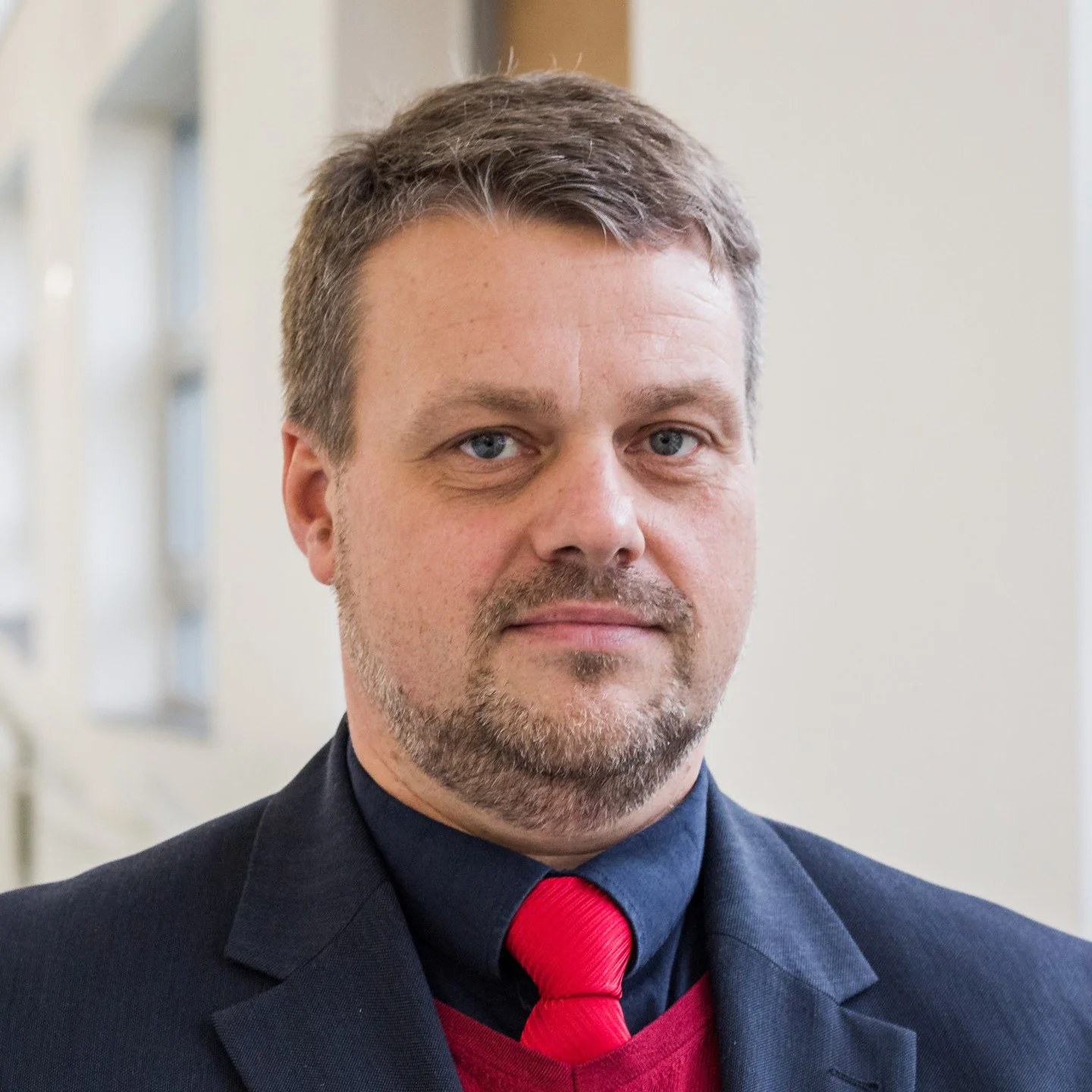
Prof. Jakub Otahal, MD, PhD
Team Leader
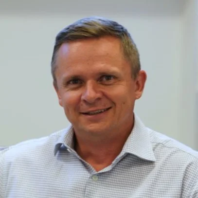
Assoc. Prof. Martin Kyncl, MD, PhD
Physician
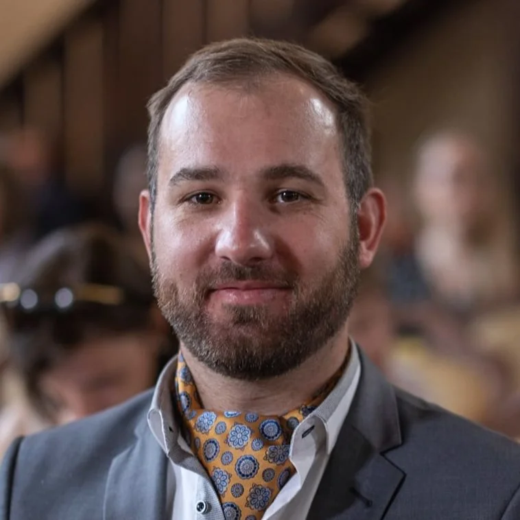
David Kala, MSc, PhD
Post-doctoral fellow

Jan Sanda
Yeva Prysiazhniuk, MSc
PhD student
Lukas Michal, MSc
PhD student

Zuzana Holubova, MD
PhD student, physician
Jan Svoboda, MSc
PhD student
Nora Profantova
Bruno Jurasek
Pavlina Pokosova, MSc
PhD student
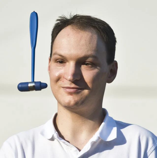
Adam Kalina, MD
Physician
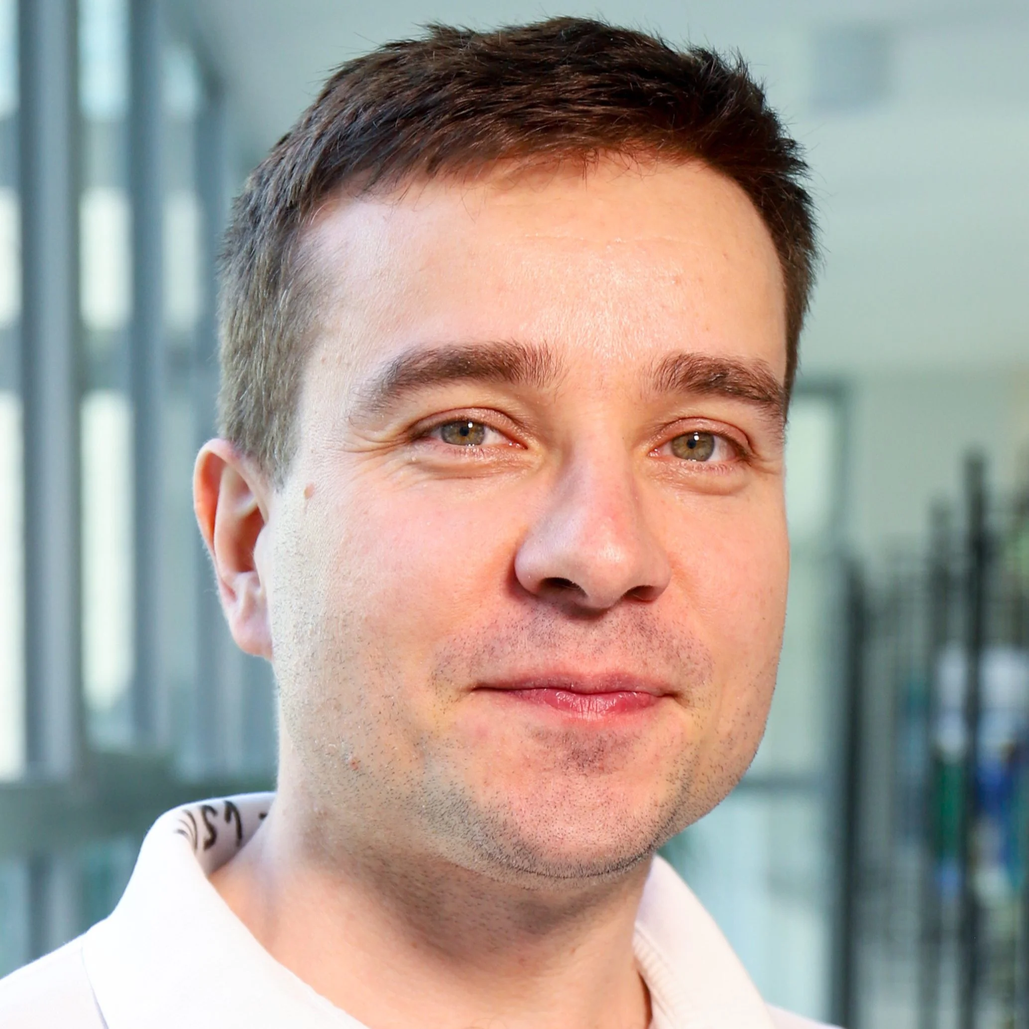
Martin Kudr, MD, PhD
Physician
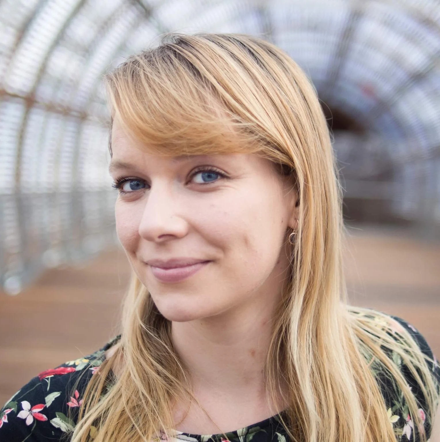
Anezka Belohlavkova, MD, PhD
Post-doctoral fellow, physician
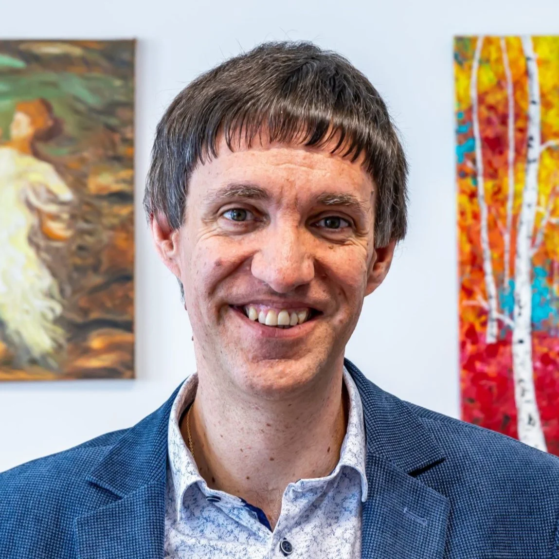
Prof. Pavel Krsek, MD, PhD
Physician
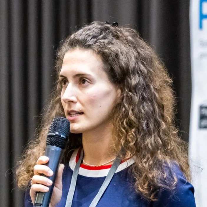
Adela Tefr Faridova, MD
PhD student
Jan Brhel, Bc DiS
Lab Technician
Veronika Borovcová
Jakub Smid, Bc
Student
Timur Abragimovich
Research areas
Acquired epilepsy
In our group we try to describe the changes that take place during the development of so-called acquired epilepsy. I focus mainly on changes in energy metabolism and cerebral perfusion. We are trying to translate the knowledge that my colleagues and I are gaining into clinical practice. We focus on improving the diagnosis of epilepsy using imaging methods, especially magnetic resonance imaging. In close collaboration with the Imaging Methods Clinic, the Neurology Clinic, and the Department of Child Neurology, we are validating for clinical practice the assessment of non-Gaussian diffusion (DKI), perfusion (DSC, pcASL) blood-brain barrier permeability (DCE, DEBBIE ASL), and newly electrical properties of tissue (MR-EPT). We are developing post-processing methods and implementing modern techniques including artificial intelligence algorithms.
Selected publications
Prysiazhniuk Y et al
Diffuse glioma molecular profiling with Arterial Spin Labeling and Dynamic Susceptibility Contrast perfusion MRI: a comparative study
Neuro-Oncology Advances 2024
Kyncl M et al
Structural MR imaging of the brain in patients with Alzheimer’s disease/ARIA assessment – initial practical information
Czech Radiology 2024
Danacikova S et al
In vitro human cell culture models in a bench-to-bedside approach to epilepsy
Epilepsia Open 2024
Buckova BR et al
Structural connectivity-based predictors of cognitive impairment in stroke patients attributable to aging
PLoS One 2023
Kyncl M et al
Recommendations for structural brain MRI in the diagnosis of epilepsy
Czech and Slovak Neurology and Neurosurgery 2023
Folbergrova J et al
Protective effect of sulforaphane on oxidative stress and mitochondrial dysfunction associated with status epilepticus in immature rats
Molecular Neurobiology 2023
Kala D et al
Evaluation of blood-brain barrier integrity by the analysis of dynamic contrast-enhanced MRI – a comparison of quantitative and semi-quantitative methods
Physiological Research 2022
Danek J et al
Sulforaphane ameliorates metabolic changes associated with status epilepticus in immature rats
Frontiers in Cellular Neuroscience 2022
Current grant projects
NU21-08-00228
Detection of changes in microstructure and structural connectivity of focal cortical dysplasias using diffusion kurtosis imaging
NU21-02-00289
Neuroimaging in prediction of haemorrhagic complications after mechanical recanalization treatment of ischemic stroke
NU23-08-00460 PediTuMRI
Advanced Magnetic Resonance Imaging of Paediatric Brain Tumors
National Recovery Program – Excelles
COGNI (NPO cooperation)
CADASIL (CERICA clinical trial)
MedWaveImage (EU-Interreg)
Microwave imaging technology transfer to innovate the medical sector
International cooperation
University of Liverpool
UMC Utrecht
HZDR Dresden
Amsterdam UMC
Fraunofer MEVIS
University of Oslo
GliMR
University of South Carolina
IKEM
Keywords
epilepsy, magnetic resonance imaging, diffusion, perfusion, energy metabolism